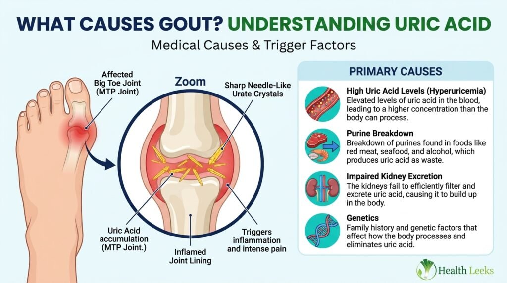
What causes gout is monosodium urate crystals triggering inflammation in a joint, after blood uric acid stays too high for too long. Food, alcohol, medicines, genes, and kidney function all influence that level, but no single meal alone creates gout.
Gout is a common form of inflammatory arthritis, known for sudden attacks. Attacks often start at night, with intense pain, swelling, redness, and tenderness in one joint.
General information only follows here, not medical advice. Fever plus a hot, inflamed joint needs urgent medical care, because joint infection can look like gout.
The Short Answer
Hyperuricemia in One View
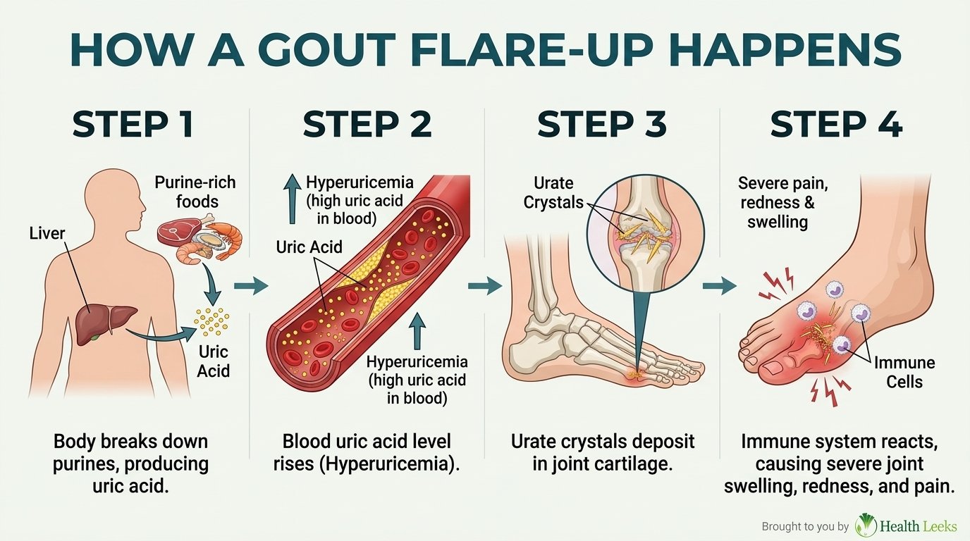
Hyperuricemia means blood uric acid above the level where crystals can form. Uric acid dissolves in blood normally, then passes through kidneys into urine.
High uric acid alone does not mean gout, and many people never develop attacks. Crystals, immune response, and joint conditions together determine whether flares occur.
Uric acid levels rise through overproduction, underexcretion, or both pathways together. Underexcretion dominates most cases, while overproduction stays less common.
Blood uric acid numbers guide clinician decisions, but never diagnose gout alone. Joint fluid microscopy, showing urate crystals, remains the gold standard for diagnosis.
Monosodium Urate Crystals in One View
Monosodium urate crystals form when uric acid stops dissolving and solidifies. Crystals are sharp, needle shaped, and deposit in joints and surrounding tissue.
Cooler joints form crystals first, because lower temperature reduces uric acid solubility. Big toe joints sit far from core warmth, with lower blood flow and temperature.
Crystals can sit silently for years before a first flare appears. Silent crystal deposits explain why a flare can seem sudden after decades of high uric acid.
Chronic crystal deposits under skin form firm, painless lumps called tophi. Tophi appear on fingers, hands, feet, elbows, and Achilles tendons in advanced gout.
Immune Inflammation in One View
Immune cells treat urate crystals as foreign bodies and attack them. Macrophages surround crystals and ingest them through phagocytosis.
Ingested crystals trigger inflammatory chemicals called cytokines, including interleukin-1 beta. Cytokines recruit more white blood cells, amplifying joint inflammation.
Inflammation causes redness, heat, swelling, and intense pain within hours. Pain often peaks within the first 8 to 12 hours after a flare starts.
Discomfort after peak pain may last from a few days to a few weeks. Joint tenderness and limited motion often linger after the worst pain eases.
How Uric Acid Is Made and Removed
Purines From the Body
Purines are natural compounds found in every cell, used to build DNA and RNA. Body cells break down constantly, releasing purines for recycling or disposal.
Purine breakdown produces hypoxanthine, then xanthine, then uric acid. Uric acid is the final waste product of purine metabolism in humans.
Cell turnover increases during illness, injury, surgery, and rapid growth. Increased turnover raises purine load, increasing uric acid production temporarily.
Body purines contribute most daily uric acid, more than food purines alone. Food matters, but internal cell turnover forms the larger baseline.
Purines From Food and Drink
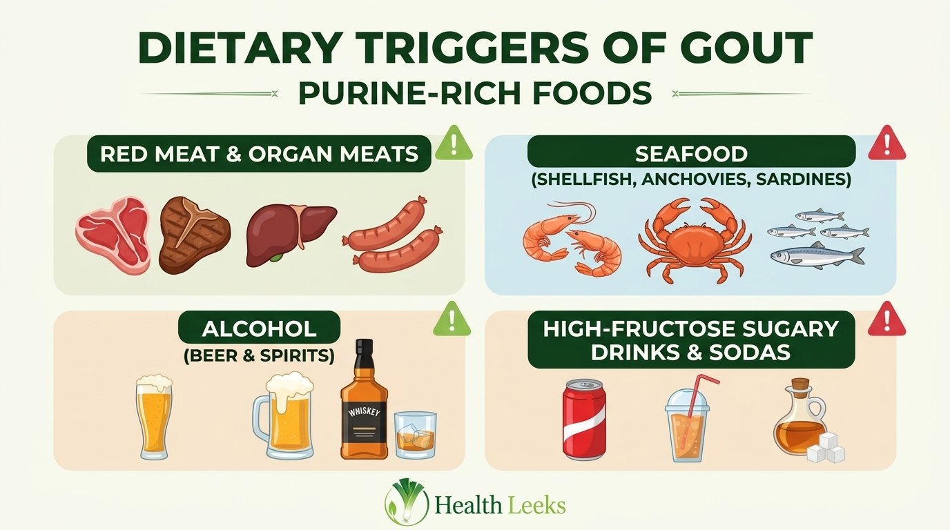
Food purines add to body purines, increasing total uric acid load. High purine foods include red meat, organ meat, and certain seafood.
Red meat includes beef, lamb, and pork, while organ meat includes liver and kidney. Game meat, including venison, goose, veal, and duck, also carries high purines.
Purine rich seafood includes anchovies, sardines, mussels, scallops, trout, and tuna. Other seafood, including cod, haddock, and herring, also appears on high purine lists.
Processed meat, including sausages and deli meat, adds purine and salt load together. Gravy and meat sauces concentrate purines from cooking meat.
Kidney Excretion
Kidneys filter uric acid from blood and excrete most of it into urine. Kidney excretion handles roughly two thirds of daily uric acid removal.
Kidney tubules reabsorb most filtered uric acid back into blood. Reabsorption balance determines how much uric acid actually leaves the body.
Kidney disease reduces filtration and excretion, raising blood uric acid. Chronic kidney disease therefore raises gout risk through underexcretion.
Dehydration concentrates blood and reduces kidney excretion efficiency. Low fluid volume makes crystal formation more likely during flares.
Gut Excretion
Gut excretion handles roughly one third of daily uric acid removal. Intestinal transporters move uric acid from blood into gut lumen for disposal.
ABCG2 transporter works in both kidney and gut for uric acid secretion. ABCG2 dysfunction reduces gut excretion, overloading kidney excretion.
Gut excretion explains why some people develop gout despite normal kidney function. Intestinal transporter variants shift excretion burden onto kidneys.
Gut bacteria and intestinal health influence uric acid handling indirectly. Research on gut excretion remains active, with transporter genetics as the clearest anchor.
Why Cooler Joints Form Crystals First
Uric acid solubility falls as temperature falls, favouring crystal formation. Peripheral joints run cooler than core organs, especially feet and hands.
Big toe joints combine cool temperature, lower blood flow, and mechanical stress. Mechanical stress from walking and footwear may further favour crystal deposits.
Ear rims also collect crystals in advanced gout, because ears run cool. Tophi on ear rims signal long standing crystal deposits, not new infection.
Knees, ankles, elbows, wrists, and fingers also develop gout flares. Any joint can flare, though big toe remains the most common first site.
Overproduction Versus Underexcretion
| Pathway | What happens | Common causes | Typical pattern |
|---|---|---|---|
| Overproduction | Body makes more uric acid than kidneys can remove | High purine diet, rapid cell turnover, rare enzyme disorders, cancer treatment | Less common, often with high urine uric acid |
| Underexcretion | Kidneys and gut remove too little uric acid | Kidney disease, dehydration, medicines, genetics, lead exposure | Dominant pathway in most cases |
| Combined | Both production rises and excretion falls | Obesity plus kidney disease, alcohol plus diuretics, diet plus genetics | Most common real world pattern |
| Normal excretion with high load | Kidneys work normally, but load overwhelms capacity | Binge alcohol, large purine meals, rapid weight loss | Flare after load spike, then level falls |
Overproduction Causes
Overproduction means the body creates uric acid faster than excretion can clear it. High purine intake contributes, but rarely explains overproduction alone.
Rapid cell turnover increases purine breakdown during cancer treatment and blood disorders. Leukemia, lymphoma, and chemotherapy increase cell death and purine load.
Psoriasis increases skin cell turnover, raising purine breakdown. Severe psoriasis therefore associates with higher uric acid levels.
Rare enzyme disorders increase purine production through metabolic errors. Phosphoribosylpyrophosphate synthetase superactivity increases purine synthesis.
Hypoxanthine-guanine phosphoribosyltransferase deficiency reduces purine recycling. Lesch-Nyhan syndrome represents the severe form of this deficiency.
Underexcretion Causes
Underexcretion means kidneys and gut remove too little uric acid. Underexcretion dominates most gout cases, even when diet looks modest.
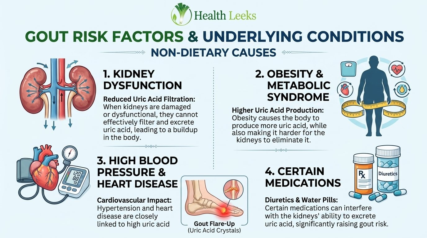
Kidney disease reduces filtration, directly reducing uric acid excretion. Chronic kidney disease therefore raises gout risk through underexcretion.
Medicines reduce uric acid excretion by blocking kidney transporters. Diuretics, low dose aspirin, and some blood pressure medicines fall in this group.
Genetic transporter variants reduce secretion or increase reabsorption. SLC2A9, ABCG2, and URAT1 variants shift excretion balance toward retention.
Lead exposure damages kidneys, causing lead nephropathy and underexcretion. Occupational and environmental lead history matters for unexplained gout.
Combined Cases
Most gout cases combine overproduction and underexcretion together. Obesity increases production while reducing excretion, creating a double effect.
Alcohol increases production and reduces excretion at the same time. Alcohol therefore combines both pathways in a single trigger.
Diet plus genetics plus kidney function together determine most risk. No single factor explains most cases, which is why blame focused on diet alone misleads.
Combined cases need clinician review of diet, medicines, conditions, and genetics together. Single factor fixes rarely control combined cases fully.
Why the Split Matters
Pathway split guides clinician decisions about testing and treatment. Urine uric acid testing helps clinicians distinguish overproduction from underexcretion in selected cases.
Overproduction cases may need production blocking treatment, while underexcretion cases may need excretion support. Treatment choice belongs to clinicians, not to articles.
Pathway knowledge also explains why diet alone sometimes fails. Underexcretion cases need more than diet changes, because kidneys drive the problem.
Patients should discuss pathway testing with clinicians, not self diagnose from food lists. Food lists alone cannot distinguish pathways reliably.
Diet Causes, With Honest Limits
Red Meat and Organ Meat
Red meat and organ meat raise uric acid through purine load. Organ meat carries the highest purine density among common meats.
Beef, lamb, and pork all contribute purines, with organ meat highest. Portion size and frequency matter more than single servings.
Mayo Clinic diuretic guidance suggests limiting meat and seafood to 6 ounces daily on a 2,000 calorie diet. Portion guidance helps patients discuss diet with clinicians, not as a prescription.
Plant proteins, low fat dairy, beans, and soy offer lower purine alternatives. Protein alternatives help patients reduce purine load without protein deficiency.
Seafood Tiers
| Seafood tier | Examples | Purine load |
|---|---|---|
| Highest | Anchovies, sardines, mussels, scallops | High |
| High | Trout, tuna, herring, mackerel, cod, haddock | High to moderate |
| Moderate | Most white fish, salmon in moderation | Moderate |
| Lower purine protein | Low fat dairy, beans, soy, eggs | Low |
Seafood purine tiers help patients prioritise reductions, not eliminate all fish. Highest tier seafood deserves the strictest limits for frequent flares.
Fish oil and omega-3 benefits do not cancel purine load for gout patients. Patients should discuss fish choices with clinicians, balancing heart and gout needs.
Shellfish and organ meat together create the highest dietary purine load. Combined high purine meals increase flare risk more than single items.
Fructose and Sugary Drinks
Fructose increases uric acid production through a distinct pathway from purines. Fructose metabolism consumes ATP, increasing purine breakdown internally.
High-fructose corn syrup in sodas and processed foods raises gout risk. Fruit flavoured drinks, non diet soda, and many processed foods carry fructose load.
Fructose from whole fruit behaves differently from added fructose in drinks. Whole fruit fibre and nutrients change absorption, though portion still matters.
Sweetened drinks deserve limits even for patients who avoid alcohol and meat. Drink sugar often explains flares in patients who eat modestly.
Purine Rich Vegetables
Purine rich vegetables, including beans, peas, lentils, and spinach, are not established gout causes in current summaries. Older purine bans included vegetables uncritically, but current guidance differs.
Vegetable purines appear less harmful than meat and seafood purines. Plant purines may behave differently due to fibre, nutrients, and overall diet pattern.
Patients should not eliminate beans, lentils, and spinach solely for gout. Nutrient loss from vegetable elimination outweighs uncertain purine benefit.
Current summaries still advise limiting meat, seafood, alcohol, and fructose drinks first. Vegetable limits belong far down the priority list, if at all.
Dairy, Coffee, and Vitamin C
Low fat dairy, coffee, and vitamin C show protective associations in observational studies. Associations mean correlation, not proven treatment effect.
Dairy proteins may increase uric acid excretion, though mechanisms remain under study. Low fat dairy suits most gout diets as a protein alternative.
Coffee drinkers show lower gout incidence in some studies, though causation remains unproven. Coffee should never replace clinician treatment for gout.
Vitamin C may increase uric acid excretion in reviews, though evidence remains mixed. Vitamin C supplements need clinician discussion, especially with kidney stones.
Alcohol Causes
Beer, Spirits, and Wine Compared
| Drink | Gout risk pattern | Mechanism |
|---|---|---|
| Beer | Highest risk, especially two or more daily | Purines from brewer’s yeast plus alcohol effects |
| Spirits | High risk | Alcohol increases production and reduces excretion |
| Wine | Lower risk than beer or spirits in studies | Alcohol effects remain, purine load lower |
| All alcohol | Risk rises with amount and binge pattern | Dehydration plus production plus excretion effects |
Beer carries purines from brewer’s yeast, adding to alcohol effects. Beer therefore combines purine load with alcohol metabolism effects.
Spirits lack beer purines, but alcohol alone raises uric acid. Spirits still increase production and reduce excretion through kidney effects.
Wine shows lower risk than beer or spirits in observational studies. Lower risk does not mean safe, especially with binge drinking or frequent use.
Amount and pattern matter more than drink type alone. Binge drinking raises risk more than occasional moderate drinking.
Production Plus Excretion Effects
Alcohol increases uric acid production through purine breakdown and lactate competition. Lactate competes with urate for kidney excretion, reducing clearance.
Alcohol also dehydrates, concentrating blood uric acid further. Dehydration plus reduced excretion creates a double risk during drinking.
Alcohol metabolism increases ATP breakdown, raising internal purine production. Internal production rises even without high purine food intake.
Alcohol effects persist into the next day, when flares often appear. Flare timing after drinking reflects crystal formation and immune response delay.
Binge Drinking and Dehydration
Binge drinking combines high alcohol load, dehydration, and sleep disruption. Each factor independently raises flare risk, together they amplify risk.
Dehydration from alcohol reduces kidney excretion and concentrates uric acid. Fluid replacement after drinking helps, but never cancels alcohol risk fully.
Holiday drinking patterns explain many seasonal gout flares. Thanksgiving, holidays, and celebrations combine alcohol, food, and dehydration.
Patients should discuss alcohol limits with clinicians, not assume abstinence alone cures gout. Alcohol reduction helps, but kidney and genetic factors remain.
Weight, Metabolic Syndrome, and Related Conditions
Obesity and Uric Acid Production
Obesity increases uric acid production and reduces kidney excretion together. Excess weight creates a double effect on uric acid balance.
Fat tissue increases purine turnover and insulin resistance. Insulin resistance reduces kidney uric acid excretion, raising blood levels.
Weight loss, done gradually with clinician guidance, lowers uric acid over time. Gradual loss helps, while rapid loss temporarily raises uric acid.
Weight management needs diet, activity, and clinician support together. Single factor weight advice rarely sustains long term uric acid control.
Hypertension, Diabetes, Heart Disease, Kidney Disease
Untreated hypertension raises gout risk through kidney effects and medicine effects. Hypertension damages kidneys over time, reducing uric acid excretion.
Diabetes and insulin resistance reduce kidney uric acid excretion. Metabolic syndrome combines obesity, hypertension, insulin resistance, and abnormal lipids.
Heart disease associates with gout through shared metabolic and kidney pathways. Gout patients face higher cardiovascular risk, requiring clinician monitoring.
Kidney disease remains the strongest condition link to gout through underexcretion. Kidney function testing belongs in gout evaluation for most patients.
Psoriasis and Blood Cell Disorders
Psoriasis increases skin cell turnover, raising purine breakdown. Severe psoriasis therefore raises uric acid production measurably.
Myeloproliferative and lymphoproliferative disorders increase cell turnover. Blood cell disorders raise purine load through rapid cell production and death.
Hemolytic anemia increases red blood cell breakdown, raising purine load. Rapid red cell turnover adds to daily purine burden.
Cancer treatment increases cell death, raising purine load during therapy. Clinicians monitor uric acid during chemotherapy for high turnover cancers.
Crash Diets and Fasting Paradox
Fasting and crash diets temporarily raise uric acid levels. Rapid weight loss increases purine breakdown and ketone competition for excretion.
Ketones compete with urate for kidney excretion during fasting. Ketoacidosis and lactic acidosis also reduce urate secretion through kidney effects.
Mayo Clinic guidance warns against fasting and quick weight loss programs for gout. Quick loss strategies temporarily raise uric acid instead of lowering it.
Gradual weight loss with adequate hydration and clinician guidance avoids the paradox. Slow loss lets excretion adapt without crystal spikes.
Medicines That Raise Uric Acid
| Medicine group | Examples | Mechanism | Action |
|---|---|---|---|
| Thiazide diuretics | Hydrochlorothiazide, chlorthalidone | Reduce urate excretion, concentrate fluid | Discuss with clinician, never stop alone |
| Loop diuretics | Furosemide, bumetanide | Reduce urate excretion, volume depletion | Discuss with clinician, never stop alone |
| Low dose aspirin | Daily low dose aspirin | Reduces urate excretion at low doses | Discuss with clinician, never stop alone |
| ACE inhibitors | Lisinopril, enalapril | May raise uric acid in some patients | Discuss with clinician, never stop alone |
| Beta blockers | Atenolol, metoprolol | May raise uric acid in some patients | Discuss with clinician, never stop alone |
| Anti-rejection drugs | Ciclosporin, tacrolimus | Reduce kidney excretion, kidney damage | Specialist guidance only, never stop alone |
| Chemotherapy agents | Various cancer treatments | Increase cell turnover and purine load | Oncology guidance only |
Thiazide and Loop Diuretics
Diuretics increase urination, lowering body fluid volume. Lower fluid volume concentrates remaining uric acid, favouring crystal formation.
Some diuretics also block kidney urate excretion directly. Reduced excretion plus concentration creates a double risk for gout.
Diuretics commonly treat hypertension, heart failure, and kidney disease. Gout risk from diuretics must balance against blood pressure and heart benefits.
Many alternative blood pressure medicines do not raise gout risk. Clinicians can discuss switching options when gout develops on diuretics.
Low Dose Aspirin
Low dose aspirin reduces urate excretion at low doses. Low dose effects differ from high dose aspirin effects on uric acid.
Low dose aspirin commonly prevents heart attack and stroke. Cardiovascular benefits often outweigh gout risk, requiring clinician judgement.
Patients should never stop low dose aspirin for gout without clinician guidance. Stopping aspirin risks heart attack and stroke, outweighing gout flare risk.
Clinicians weigh gout frequency against cardiovascular risk for each patient. Individual risk balance determines aspirin continuation decisions.
ACE Inhibitors and Beta Blockers
ACE inhibitors and beta blockers may raise uric acid in some patients. Mechanisms vary, with kidney effects and metabolic effects both contributing.
ACE inhibitors protect kidneys and heart in many patients. Kidney protection benefits may outweigh modest uric acid increases.
Beta blockers control heart rate and blood pressure effectively. Heart benefits often outweigh gout risk for cardiac patients.
Medicine review should consider all medicines together, not single drugs alone. Combined medicine effects matter more than single drug effects.
Anti-Rejection Drugs
Anti-rejection drugs prevent organ rejection after transplant. Ciclosporin and tacrolimus reduce kidney function and urate excretion.
Transplant patients face high gout risk from medicines plus kidney function. Specialist guidance manages gout without risking organ rejection.
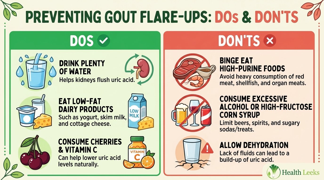
Anti-rejection drugs must never stop for gout without specialist guidance. Stopping risks organ loss, far outweighing gout flare harm.
Transplant gout management needs rheumatology plus transplant team coordination. Coordinated care balances gout control with organ protection.
Never Stop a Medicine Without Clinician Guidance
Stopping blood pressure, heart, aspirin, or anti-rejection medicines risks serious harm. Heart attack, stroke, kidney failure, and organ rejection outweigh gout flares.
Medicine review appointments should include full medicine lists, including over the counter drugs. Over the counter aspirin and diuretics also affect uric acid.
Clinicians can often switch medicines to lower gout risk safely. Switching requires monitoring, not abrupt stopping.
Patients should bring gout history to every medicine review. Gout history changes medicine choices for hypertension and heart disease.
Genetics and Family History
Heritability Ranges
Serum urate heritability estimates range from 45 to 73 percent in reviews. Heritability means genetic variation explains much of uric acid variation between people.
Gout heritability estimates around 30 percent in people of European descent. Gout heritability runs lower than urate heritability, because flares need crystals plus immune response.
Heritability does not mean destiny, because environment and medicines modify risk. Genes set baseline excretion, while diet, alcohol, and medicines shift levels further.
Family history raises gout risk even without genetic testing. Family history reflects shared genes, diet, and environment together.
SLC2A9, ABCG2, URAT1
| Gene | Protein | Effect on uric acid |
|---|---|---|
| SLC2A9 | GLUT9 transporter | Variants increase reabsorption or reduce excretion, raising urate |
| ABCG2 | ABCG2 transporter, kidney and gut | Q141K variant reduces transport by about 50 percent, raising urate |
| SLC22A12 | URAT1 transporter, kidney | Main reabsorption transporter, variants shift excretion balance |
| SLC17A1 | NPT1 transporter | Variants affect urate secretion, with smaller effects |
| PDZK1 | Scaffolding protein | Variants affect transporter organisation, with smaller effects |
SLC2A9 explains about 3 percent of urate variance, large for a single gene. SLC2A9 isoforms differ by kidney side, affecting reabsorption versus excretion.
ABCG2 Q141K variant reduces urate transport by about 50 percent in functional studies. Reduced gut excretion overloads kidney excretion, raising blood urate.
URAT1 represents the main kidney reabsorption transporter for urate. URAT1 variants shift reabsorption balance, affecting excretion efficiency.
Transporter variants also affect responses to fructose and medicines variably. Genetic effects on diet and medicine responses remain mixed in studies.
Rare Enzyme Disorders
Familial juvenile hyperuricemic nephropathy causes early kidney disease and gout. Early onset gout with kidney disease suggests familial evaluation.
Medullary cystic kidney disease causes kidney cysts and hyperuricemia. Kidney cysts plus gout in young patients warrants specialist evaluation.
Phosphoribosylpyrophosphate synthetase superactivity increases purine production. Increased production causes early, severe gout in affected families.
Hypoxanthine-guanine phosphoribosyltransferase deficiency reduces purine recycling. Lesch-Nyhan syndrome represents severe deficiency with gout and neurological features.
Ancestry and Prevalence Differences
Prevalence varies by ancestry, with higher rates in some populations. African American, Maori, Japanese, and Hmong populations show higher hyperuricemia or gout rates in studies.
Arthritis Foundation 2026 infographic reports 9.2 million United States adults with gout. Older CDC based estimates report 8.3 million adults and 3.9 percent prevalence.
North America review reports 5.1 percent United States prevalence, with different methods and dates. Prevalence figures conflict by source, method, and date, so never merge them.
Black adults face 46 percent higher gout likelihood than white adults in Arthritis Foundation data. Disparities reflect genetics, kidney disease, access, and social determinants together.
Age, Sex, and Hormones
Men 30 to 50
Men develop gout most often between ages 30 and 50. Male uric acid levels run higher than female levels before menopause.
Male risk rises with age, obesity, alcohol, and metabolic conditions. Age plus metabolic factors together drive male gout incidence.
Young men with gout often show family history or early kidney issues. Early onset gout warrants clinician evaluation for genetics and kidney function.
Men face gout about three times more often than premenopausal women in Arthritis Foundation data. Sex differences narrow after menopause as female urate levels rise.
Women After Menopause
Women develop gout most often after age 60, following menopause. Postmenopausal uric acid levels approach male levels as estrogen falls.
Estrogen promotes kidney uric acid excretion before menopause. Estrogen loss after menopause reduces excretion, raising blood urate.
Postmenopausal women with gout often use diuretics or have kidney disease. Medicine and kidney factors combine with hormone changes after menopause.
Women with gout face underdiagnosis, because gout stereotypes focus on men. Clinicians should consider gout in postmenopausal women with sudden joint pain.
Estrogen and Uric Acid
Estrogen increases kidney uric acid excretion through transporter effects. Higher excretion keeps premenopausal uric acid levels lower.
Pregnancy and hormone therapy change estrogen levels and uric acid handling. Hormone changes need clinician discussion, not self adjustment.
Estrogen loss explains much of the postmenopausal gout rise. Hormone effects combine with age, weight, and kidney function changes.
Hormone therapy for gout prevention remains a clinician decision, not a self treatment. Risks and benefits vary by individual history.
Flare Triggers, Distinct From Underlying Cause
| Trigger | How it triggers a flare | Timing |
|---|---|---|
| Dehydration | Concentrates uric acid, reduces excretion | Hours to days after fluid loss |
| Alcohol binge | Increases production, reduces excretion, dehydrates | Next day or days after drinking |
| Large purine meal | Increases purine load temporarily | Hours to days after meal |
| Fructose drink load | Increases internal purine production | Hours after large fructose intake |
| Surgery | Tissue stress, cell turnover, dehydration, immobility | Days after surgery |
| Injury | Tissue damage, inflammation, cell turnover | Days after injury |
| Illness and infection | Cell turnover, dehydration, medicine changes | During or after illness |
| Crash diet or fasting | Ketone competition, purine breakdown | Days into fasting or rapid loss |
| Starting urate-lowering therapy | Crystal mobilisation during treatment start | Weeks after starting, clinician monitored |
| Stress | Sleep disruption, dehydration, behaviour changes | Variable, often with other triggers |
Dehydration
Dehydration concentrates blood uric acid and reduces kidney excretion. Heat, illness, vomiting, diarrhoea, and alcohol all cause dehydration.
Adequate hydration supports kidney excretion and crystal prevention. Hydration needs vary by kidney disease, heart disease, and medicine use.
Hydration alone never cures gout, but dehydration often triggers flares. Fluid balance matters most during heat, illness, and alcohol use.
Patients with heart or kidney disease need individual fluid guidance. Fluid limits for heart failure conflict with gout hydration advice, requiring clinician balance.
Alcohol and Food Load
Large alcohol plus food loads combine purine, fructose, and dehydration effects. Holiday meals combine all three triggers in one evening.
Single large meals rarely cause gout without underlying hyperuricemia. Underlying crystal deposits plus load spike together trigger flares.
Food load triggers matter most for patients with existing crystal deposits. Crystal deposits plus load spike create immune response within hours.
Trigger avoidance helps, but underlying crystal burden drives flare frequency. Long term control needs crystal dissolution, not only trigger avoidance.
Surgery, Injury, Illness, and Stress
Surgery stresses the body through tissue damage, cell turnover, and dehydration. Postoperative immobility and medicine changes add further risk.
Injury causes local inflammation and cell turnover near joints. Joint injury can trigger flares in joints with existing crystal deposits.
Illness increases cell turnover, dehydration, and medicine changes. Fever, infection, and hospitalisation often precede flares.
Stress disrupts sleep, hydration, diet, and medicine adherence. Stress alone rarely causes gout, but stress behaviours trigger flares.
Starting Urate-Lowering Therapy
Starting urate-lowering therapy can trigger flares during early treatment. Crystal mobilisation as urate levels fall can provoke immune response.
Flare risk during treatment start does not mean treatment failed. Clinicians often use flare prevention strategies during early treatment months.
Patients should never stop urate-lowering therapy for a flare without clinician guidance. Stopping restarts crystal accumulation and prolongs control time.
Treatment start flares decrease over time as crystal burden dissolves. Long term control reduces flare frequency as deposits clear.
Trigger Diary
- Date and time: Record flare start, peak pain time, and duration.
- Joint: Record which joint flared, pain severity, redness, and heat.
- Food and drink: Record alcohol, meat, seafood, and fructose drinks in prior 48 hours.
- Fluid: Record water intake, heat exposure, illness, vomiting, or diarrhoea.
- Medicines: Record new medicines, stopped medicines, diuretics, and aspirin use.
- Weight: Record rapid weight changes, fasting, or crash dieting.
- Surgery and injury: Record recent surgery, injury, illness, or stress events.
- Treatment: Record urate-lowering therapy start, dose changes, and clinician advice.
Gout Versus Pseudogout Versus Septic Arthritis
| Feature | Gout | Pseudogout | Septic arthritis |
|---|---|---|---|
| Crystal | Monosodium urate | Calcium pyrophosphate dihydrate | No crystal, bacterial infection |
| Typical joints | Big toe, foot, ankle, knee | Knee, wrist, shoulder, hip | Any joint, often knee, hip, shoulder |
| Typical age | Men 30 to 50, women after menopause | Older adults, often over 60 | Any age, with risk factors |
| Diet trigger | Purines, alcohol, fructose | No diet trigger | No diet trigger |
| Metabolic causes | Hyperuricemia, kidney disease, metabolic syndrome | Thyroid disease, iron overload, high calcium, low magnesium | Infection, immune suppression, joint damage |
| Diagnosis | Joint fluid microscopy showing urate crystals | Joint fluid microscopy showing calcium pyrophosphate crystals | Joint fluid culture and microscopy, urgent evaluation |
| Urgency | Urgent if fever plus hot joint, otherwise prompt | Prompt evaluation, usually not emergency | Emergency, joint infection can destroy joint quickly |
Gout Versus Pseudogout
Gout and pseudogout both cause crystal arthritis with pain, swelling, and redness. Crystal type distinguishes them, not symptoms alone.
Pseudogout crystals form from calcium pyrophosphate in cartilage. Cartilage calcification, known as chondrocalcinosis, often accompanies pseudogout.
Pseudogout lacks purine, alcohol, and fructose triggers. Diet changes that help gout do not help pseudogout.
Pseudogout associates with older age, genetics, thyroid disease, and mineral disorders. Iron overload, high calcium, and low magnesium all associate with pseudogout.
Gout Versus Septic Arthritis
Septic arthritis means bacterial infection inside a joint, requiring emergency care. Infection can destroy joints quickly without treatment.
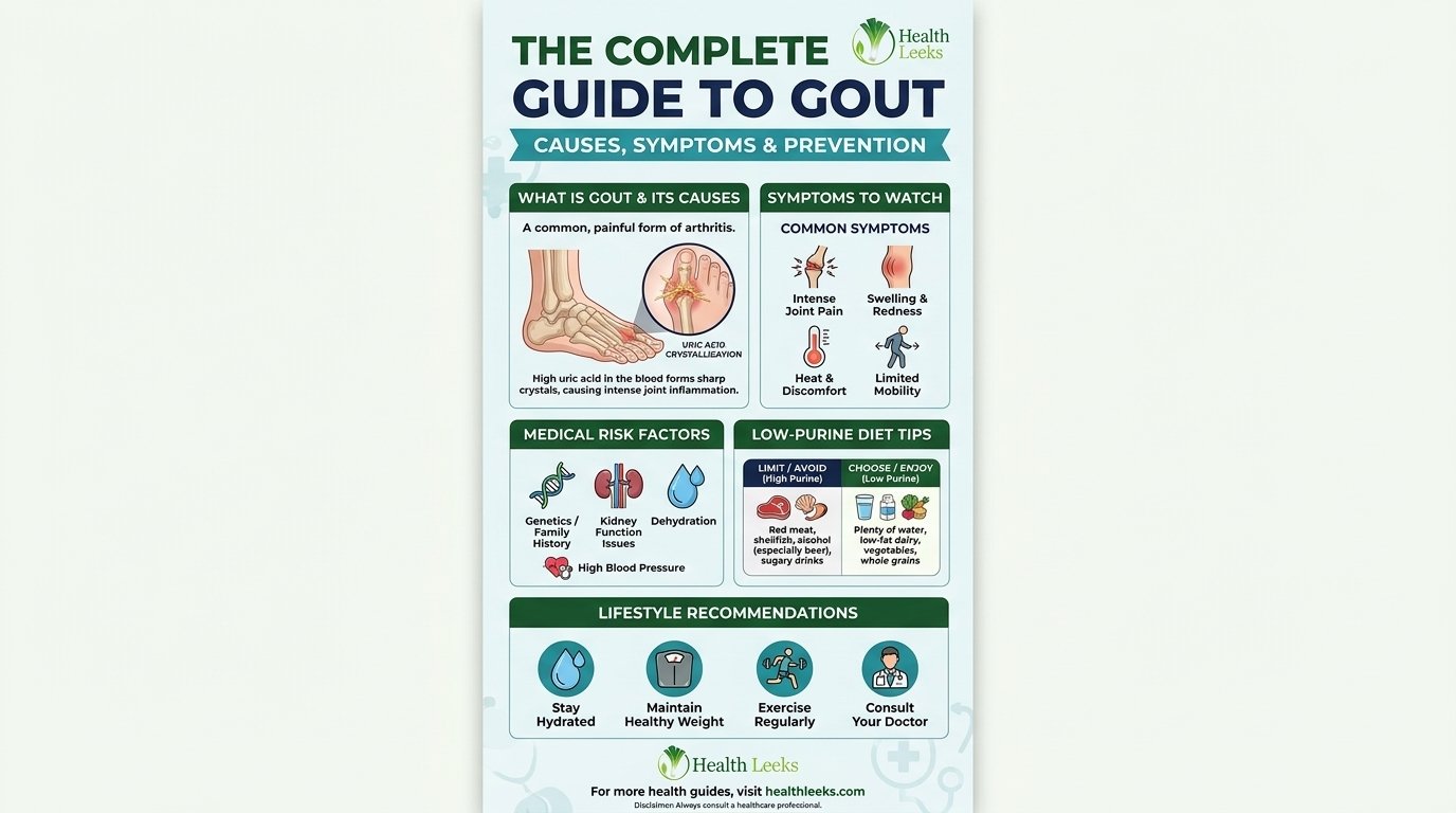
Fever plus a hot, inflamed joint signals possible infection, even with gout history. Gout history does not rule out infection, and both can coexist.
Joint fluid culture and microscopy distinguish infection from crystal arthritis. Culture results guide antibiotics, while microscopy guides crystal diagnosis.
Patients should seek urgent care immediately for fever plus hot joint. Urgent evaluation prevents joint destruction and bloodstream infection.
Stages and Complications
Asymptomatic Hyperuricemia
Asymptomatic hyperuricemia means high uric acid without gout symptoms. Most people with high uric acid never develop gout.
Asymptomatic hyperuricemia needs monitoring, not automatic treatment. Clinicians weigh level, kidney function, and risk factors before treating.
Crystal deposits can form silently during asymptomatic hyperuricemia. Silent deposits explain why first flares seem sudden after years of high levels.
Lifestyle and medicine review help asymptomatic hyperuricemia patients. Diet, alcohol, weight, and medicine review reduce future flare risk.
First Flare
First flares often affect one joint, most often the big toe. First flares start suddenly, often at night, with intense pain.
First flare pain peaks within 8 to 12 hours, then eases over days. Discomfort may linger for days to weeks after peak pain.
First flare evaluation includes joint examination, blood tests, and joint fluid testing when possible. Joint fluid testing confirms crystals and rules out infection.
Single flare does not always mean lifelong treatment. Clinicians weigh recurrence risk, kidney function, and crystal burden before long term therapy.
Recurrent Gout
Recurrent gout means multiple flares per year, with increasing frequency over time. Untreated gout tends toward more frequent flares.
Recurrent flares damage joints through repeated inflammation. Joint damage accumulates with each flare, reducing range of motion.
Recurrent gout needs long term urate control, not only flare treatment. Flare treatment alone never dissolves crystal deposits fully.
Clinicians discuss urate-lowering therapy when flares recur frequently. Treatment decisions weigh flare frequency, tophi, kidney stones, and kidney function.
Chronic Tophaceous Gout
Chronic tophaceous gout means persistent crystal deposits under skin and in joints. Tophi form firm, painless lumps in advanced, untreated gout.
Tophi can become swollen and tender during flares. Tophi also limit joint motion and damage surrounding tissue over time.
Tophi signal long standing hyperuricemia and need long term control. Crystal dissolution takes months to years of sustained urate control.
Tophi reduction requires sustained serum urate below clinician target levels. Clinician targets often sit below 6 mg per dL for crystal dissolution.
Kidney Stones
Urate crystals can collect in urinary tracts, causing kidney stones. Kidney stones cause flank pain, blood in urine, and urinary blockage.
Kidney stones associate with high urine uric acid and low urine volume. Dehydration and high purine load increase stone risk.
Kidney stone prevention includes hydration, diet review, and urate control. Stone history changes gout management priorities for clinicians.
Kidney stones need urgent care for severe pain, fever, or urinary blockage. Blocked urine flow can damage kidneys quickly without treatment.
How Gout Is Diagnosed, Briefly
Joint Fluid Microscopy
Joint fluid aspiration removes fluid from an affected joint for microscopy. Microscopy shows needle shaped urate crystals under polarised light.
Joint fluid testing works best soon after flare onset. Crystals dissolve over time, making later testing less reliable.
Ultrasound guidance helps clinicians aspirate small or difficult joints. Ultrasound also shows crystal deposits and joint inflammation.
Joint fluid culture runs alongside microscopy when infection remains possible. Culture distinguishes septic arthritis from crystal arthritis definitively.
Blood Uric Acid Limits
Blood uric acid testing measures serum urate at one moment in time. Levels fluctuate with diet, alcohol, medicines, illness, and flare status.
High uric acid does not diagnose gout, because most high levels never cause flares. Low uric acid during a flare does not rule out gout either.
Blood testing guides treatment and monitoring, not diagnosis alone. Clinicians use blood levels to track urate control over time.
Patients should discuss blood test timing with clinicians, especially during flares. Flare status affects blood urate interpretation.
Ultrasound, Dual-Energy CT, X-Ray Limits
Ultrasound shows crystal deposits and joint inflammation noninvasively. Ultrasound helps when joint fluid aspiration proves difficult.
Dual-energy CT distinguishes urate deposits from calcium deposits. Dual-energy CT helps experienced radiologists identify crystal burden.
X-ray shows bone erosion and calcifications only in advanced disease. X-ray cannot diagnose early gout, because early crystals remain invisible.
Imaging supplements, never replaces, joint fluid testing for diagnosis. Imaging plus clinical history guides diagnosis when aspiration fails.
What Does Not Cause Gout
One Meal Alone
One meal alone rarely causes gout without underlying hyperuricemia. Underlying crystal deposits plus load spike together trigger flares.
Blaming one meal ignores genetics, kidney function, and long term urate levels. Single meal blame creates stigma without improving control.
Diet matters, but diet alone explains only part of gout risk. Reviews attribute about 12 percent of gout to dietary factors in some estimates, though methods vary.
Long term patterns matter more than single meals for gout control. Consistent diet, alcohol, and weight patterns shape urate levels over years.
Purine Rich Vegetables Alone
Purine rich vegetables alone do not cause gout in current summaries. Beans, peas, lentils, and spinach carry purines, but evidence for harm remains weak.
Vegetable elimination for gout risks nutrient loss without clear benefit. Plant proteins support overall health and metabolic control.
Patients should prioritise meat, seafood, alcohol, and fructose limits first. Vegetable limits belong far down the priority list.
Cold Weather Alone
Cold weather alone does not cause gout, though cooler joints favour crystals. Cold exposure may trigger flares in joints with existing deposits.
Seasonal flares often reflect holidays, alcohol, food, and dehydration together. Winter holidays combine all triggers, not cold alone.
Warmth helps joint comfort during flares, but never treats crystal burden. Warm compresses soothe symptoms without dissolving crystals.
Contagion
Gout is not contagious and cannot spread person to person. Gout results from internal uric acid handling, not infection.
Family clustering reflects shared genes and environment, not contagion. Shared diet and genetics explain family patterns, not transmission.
Patients need not isolate for gout, though flares limit mobility. Mobility limits reflect pain, not infection risk to others.
Frequently Asked Questions
What causes gout?
Gout is caused by monosodium urate crystals triggering inflammation in a joint. Crystals form when blood uric acid stays too high, a state called hyperuricemia.
Uric acid comes from purine breakdown in the body and in food. Kidneys excrete most uric acid, while gut excretion handles the remainder.
What causes a gout flare?
Flares occur when crystals trigger immune inflammation, often after a trigger spike. Triggers include dehydration, alcohol, large purine meals, surgery, injury, and illness.
Underlying crystal deposits must exist before triggers can cause flares. Trigger avoidance helps, but crystal burden drives flare frequency.
What causes high uric acid?
High uric acid results from overproduction, underexcretion, or both pathways. Kidney disease, medicines, genetics, diet, alcohol, and obesity all raise levels.
Body purine breakdown contributes most daily uric acid, more than food alone. Food, alcohol, and fructose add to internal production.
What are uric acid crystals?
Uric acid crystals are sharp, needle shaped monosodium urate deposits. Crystals form when uric acid stops dissolving and solidifies in joints.
Crystals trigger immune cells to release inflammatory chemicals, causing pain. Crystal deposits can sit silently for years before flares.
How do uric acid crystals cause pain?
Immune cells ingest crystals and release cytokines, including interleukin-1 beta. Cytokines recruit more white blood cells, amplifying inflammation.
Inflammation causes redness, heat, swelling, and intense pain within hours. Pain often peaks within 8 to 12 hours after flare onset.
Does alcohol cause gout?
Alcohol raises gout risk through production, excretion, and dehydration effects. Beer carries the highest risk, spirits next, wine lower but still risky.
Binge drinking raises risk more than occasional moderate drinking. Amount and pattern matter more than drink type alone.
Does beer cause gout more than wine?
Beer shows higher gout risk than wine in observational studies. Beer purines from brewer’s yeast add to alcohol effects.
Wine still raises risk, especially with binge drinking or frequent use. Lower risk does not mean safe for gout patients.
Does red meat cause gout?
Red meat raises uric acid through purine load, especially organ meat. Beef, lamb, pork, and organ meat all contribute purines.
Portion size and frequency matter more than single servings. Plant proteins offer lower purine alternatives for regular meals.
Does seafood cause gout?
Seafood raises uric acid through purine load, especially anchovies and sardines. Mussels, scallops, trout, and tuna also carry high purines.
Highest tier seafood deserves strictest limits for frequent flares. Moderate fish choices need clinician discussion for heart health balance.
Does sugar cause gout?
Added sugar, especially fructose, raises uric acid through internal purine production. Fructose metabolism consumes ATP, increasing purine breakdown.
High-fructose corn syrup in sodas and processed foods raises gout risk. Whole fruit behaves differently from added fructose in drinks.
Does dehydration cause gout?
Dehydration concentrates blood uric acid and reduces kidney excretion. Heat, illness, vomiting, diarrhoea, and alcohol all cause dehydration.
Adequate hydration supports kidney excretion, though hydration alone never cures gout. Heart and kidney disease patients need individual fluid guidance.
Does obesity cause gout?
Obesity increases uric acid production and reduces kidney excretion together. Fat tissue increases purine turnover and insulin resistance.
Gradual weight loss with clinician guidance lowers uric acid over time. Rapid weight loss temporarily raises uric acid, worsening flares.
Does kidney disease cause gout?
Kidney disease reduces uric acid excretion, raising blood uric acid. Chronic kidney disease therefore raises gout risk through underexcretion.
Kidney function testing belongs in gout evaluation for most patients. Kidney disease and gout worsen each other without coordinated care.
Do diuretics cause gout?
Diuretics raise gout risk by concentrating fluid and reducing urate excretion. Thiazide and loop diuretics both affect kidney urate handling.
Patients must never stop diuretics for gout without clinician guidance. Clinicians can discuss switching blood pressure medicines safely.
Does aspirin cause gout?
Low dose aspirin reduces urate excretion at low doses. Low dose aspirin commonly prevents heart attack and stroke.
Patients must never stop low dose aspirin for gout without clinician guidance. Cardiovascular benefits often outweigh gout risk for individual patients.
Can surgery cause gout?
Surgery can trigger gout through tissue stress, cell turnover, dehydration, and immobility. Postoperative flares often appear days after surgery.
Patients with gout history should tell surgical teams before operations. Surgical teams can plan hydration, medicine review, and flare prevention.
Can fasting cause gout?
Fasting and crash diets temporarily raise uric acid levels. Ketones compete with urate for kidney excretion during fasting.
Gradual weight loss with adequate hydration avoids the fasting paradox. Quick loss strategies temporarily raise uric acid instead of lowering it.
Is gout genetic?
Gout shows strong genetic influence, with serum urate heritability from 45 to 73 percent in reviews. SLC2A9, ABCG2, and URAT1 variants shift excretion balance.
Genes set baseline excretion, while diet, alcohol, and medicines shift levels further. Family history raises risk even without genetic testing.
Can gout happen without alcohol?
Gout frequently happens without alcohol, through genetics, kidney disease, and medicines. Alcohol explains only part of gout risk overall.
Non drinkers with gout often show family history, kidney disease, or diuretic use. Clinicians should evaluate non alcohol causes thoroughly.
Can gout happen with normal uric acid?
Gout can occur during a flare even when blood uric acid looks normal. Levels fluctuate with diet, alcohol, medicines, illness, and flare status.
Normal blood uric acid during a flare does not rule out gout. Joint fluid microscopy remains the gold standard for diagnosis.
Can high uric acid happen without gout?
Most people with high uric acid never develop gout. Crystal formation plus immune response together determine whether flares occur.
Asymptomatic hyperuricemia needs monitoring, not automatic treatment. Clinicians weigh level, kidney function, and risk factors before treating.
Why does gout affect the big toe first?
Big toe joints run cooler, with lower blood flow and mechanical stress. Cooler temperature reduces uric acid solubility, favouring crystal formation.
Mechanical stress from walking and footwear may further favour deposits. Big toe combination of coolness, flow, and stress explains first flare site.
What is the difference between gout and pseudogout?
Gout uses monosodium urate crystals, while pseudogout uses calcium pyrophosphate crystals. Pseudogout lacks purine, alcohol, and fructose triggers.
Pseudogout typically affects knees, wrists, and shoulders in older adults. Diagnosis requires joint fluid microscopy to distinguish crystal types.
When should I seek urgent care for a hot joint?
Seek urgent care immediately for fever plus a hot, inflamed joint. Fever plus hot joint signals possible joint infection, even with gout history.
Joint infection can destroy joints quickly without treatment. Urgent joint fluid culture and microscopy distinguish infection from gout.
Conclusion
What causes gout is crystal inflammation from sustained hyperuricemia, shaped by kidney excretion, gut excretion, genetics, diet, alcohol, medicines, and related conditions. No single food, drink, or habit alone explains most cases, and no single trigger list alone controls gout.
Track underlying causes and flare triggers separately, with a trigger diary and full medicine list. Discuss diet, alcohol, weight, kidney function, and medicine options with a clinician, never stopping prescribed medicines alone, and seek urgent care for fever plus a hot joint.
Choose the next step that fits your history: clinician review after a first flare, trigger diary for recurrent flares, and long term urate discussion for chronic gout. Crystal burden dissolves only with sustained control, so early evaluation, honest cause review, and steady follow up matter more than blame, single food bans, or quick fixes.